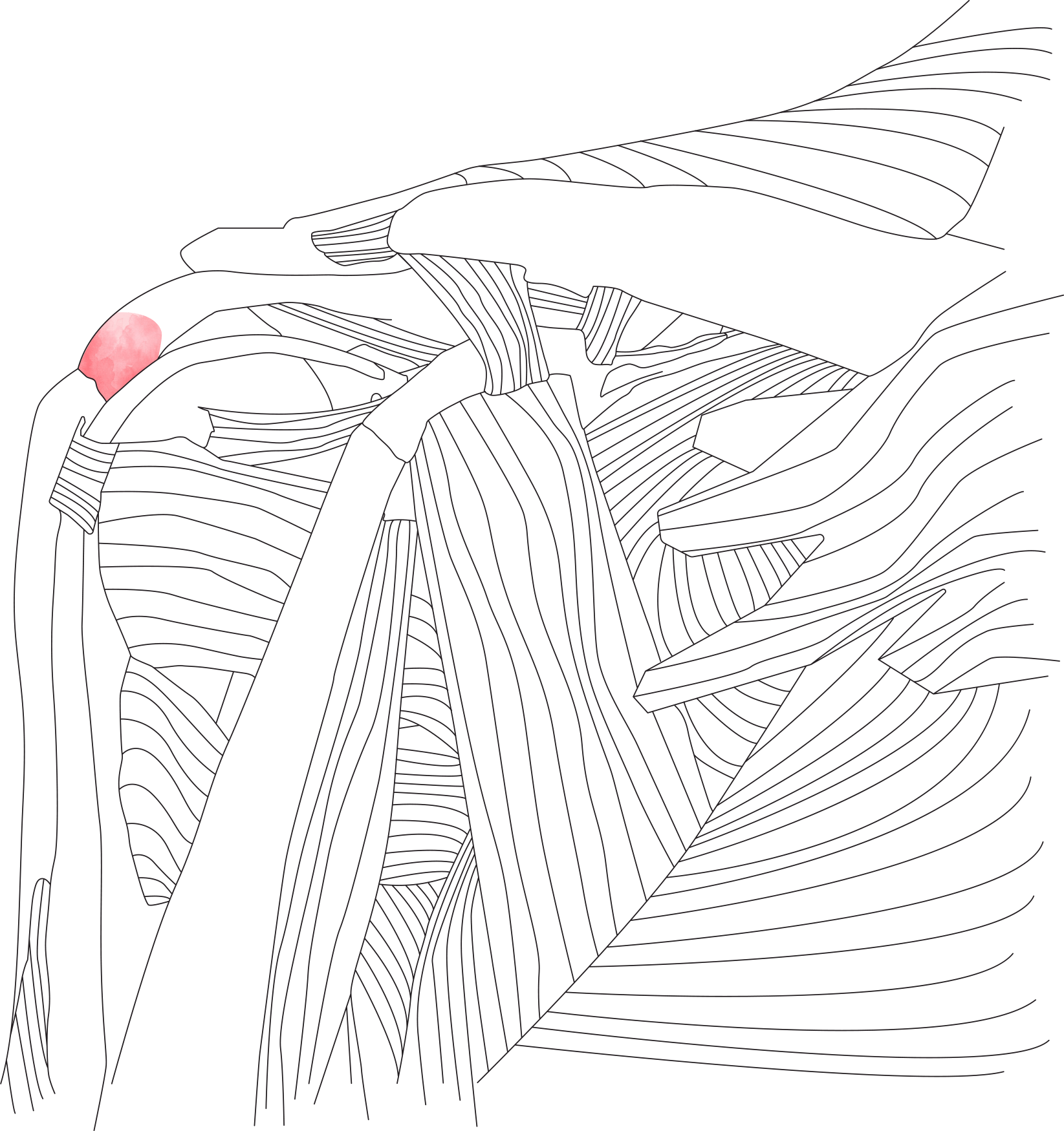
Calcific Tendonitis is a relatively common shoulder condition. It has a peak onset age of 40 years of age. It affects women more commonly than men. The most commonly affected of the rotator cuff tendons is the supraspinatus tendon.
It seems that the presence of calcium in the rotator cuff tendons is relatively common. Some studies have suggested that between 2 to 20% of people may have calcium present in their rotator cuff tendons without symptoms. The shoulder becomes symptomatic when an inflammatory reaction occurs in relation to the calcium within the rotator cuff tendon. It is the inflammatory reaction rather than the presence of the calcium that causes the symptoms.
There are two theories as to how calcium is deposited in the tendon. One theory suggests that degenerate areas of tendon act as a focus for the calcium to form within the tendon. The other theory claims that calcium is deposited within areas of tendon with low blood flow and a degree of hypoxia.
The argument against the degenerate tendon theory is that calcium is in fact more commonly seen in more normal looking tendon rather than tendons with significant tears.
Several stages of calcific tendonitis are described. There appears to be some initial change in the fibrocartilage cells within the tendon that allows calcium to be deposited within the tendon. This precalcific stage is followed by a calcific formative stage in which chalky calcium deposits form. This stage tends to be a chronic, painless stage. This is different to the resorptive stage, in which the body mounts an inflammatory reaction to the calcium. Macrophages from the blood stream react to the calcium and start to reabsorb the calcium deposit. The calcium lump becomes soft and toothpaste like due to the enzymes released by the macrophages. This stage is usually acutely painful.
Patients with calcium in their rotator cuff tendons may be entirely asymptomatic. They may present with mild to moderate chronic pain and discomfort in their shoulder or they may develop acute severe pain in the shoulder related to the inflammatory reaction against the calcium deposit as the body tries to reabsorb it.
Pain is typically felt down the side of the upper arm. The shoulder tends to ache at night time. Pain is often felt as the arm is lifted overhead with the swollen area of tendon catching on the under surface of the acromion and impinging.
X-Rays shows the deposited calcium very well. However, several views will need to be taken to be certain to demonstrate it. X-Rays need to be taken with the arm in neutral rotation and in internal and external rotation.
Ultrasound is particularly sensitive to calcium and will invariably demonstrate it.
A MRI examination is not very good at demonstrating calcium deposits because the calcium shows up dark on the scans, as does the tendon in which the calcium is sitting in. So, it is often hard to distinguish the calcium lump within the tendon because both appear dark on the MRI scans.


